Plain Radiography To Evaluate Severity
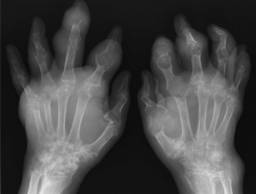
A recent study has identified a valid radiographic damage index in chronic gout. Such a scoring system is required for future studies to determine the impact of intensive urate-lowering on radiographic damage, and to guide therapeutic decision-making in patients with chronic gout. The gout radiographic damage index is a modified Sharp/van der Heijde scoring method of erosion and joint space narrowing, which incorporates the joints scored in the rheumatoid arthritis scoring method and the hand distal interphalangeal joints. This index was shown to be reproducible, feasible, and able to discriminate between early and late disease . This damage index has shown to be strongly associated with functional capacity .
A comparison of plain radiography and computed tomography has shown very good agreement between the two methods for assessment of gouty erosions, providing further validation for the erosion component of the gout radiographic damage index . Some mismatch was evident, however, with higher radiographic erosion scores at the distal interphalangeal joints. It is probable that assessment of these joints for erosion by plain radiography may be less reliable due to their small size, and due to the presence of concurrent degenerative joint disease.
Can You Remove Gout
It ought to be fairly apparent why youd want to get gone gout, but is it possible in fact?
Sure will be, but theres not a one-size matches all solution.
Within the next section, well be going over whats worked best for us!
You wont want to lose out on this free video tutorial.
NOTICE: Id highly recommend going to your doctor or seeing a specialist about this situation, since we arent experts. See our medical disclaimer for more details.
We dont know what will work for you, but we know whats worked for us and others
Who Needs A Test
PsA can affect anyone. It develops most frequently in young adults, but it can appear at any age.
People with psoriasis or a family history of psoriasis are more likely to develop PsA and should be aware of the symptoms.
If someone develops arthritis symptoms and has either psoriasis or a family history of it, they should ask a doctor whether their symptoms could result from PsA or another autoimmune inflammatory disorder.
Read Also: Is Onions Good For Gout
Monitoring Disease Activity And Damage
Plain XR provides a very blunt imaging instrument with which to try to track the progress of joint damage in gout and its response to therapy. McCarthy and colleagues studied a group of 39 patients for 10 years and found no correlation between XR changes and serum urate concentration, and this suggests that XR may not be sufficiently sensitive to monitor change in bony damage over this time frame. More recently, a specific gout radiographic scoring method has been developed and validated and may improve sensitivity to change in longitudinal studies . With the development of powerful and often costly urate-lowering therapies, the focus has shifted to the possibility that advanced imaging could be useful in this context, providing sensitivity to change over a shorter timeframe that would be clinically relevant. Of these modalities, MRI and CT have the facility to allow storage of standardized digital images and so are particularly suitable for use in longitudinal studies.
Symptoms And Signs Of Gout In Foot

An attack of gout is often sudden. Symptoms:
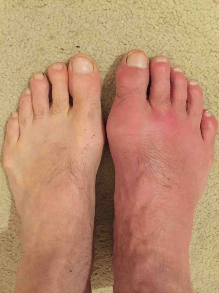
- It may present with excruciatingly painful swelling of joints in the big toe, it is known as Podagra. The joint may be stiff and appear red or purple, very swollen, and tender to even light touch. Other gout sites include the instep, wrist, ankle, fingers, and knee.
- Skin may peel and itch as healing begins.
- An attack often begins at night the acute phase lasts up to 12 hours. If untreated, the inflammation may last up to two weeks. In 10 percent of people, acute episodes present in more than one joint.
- Kidney stones precede the onset of gout in 14 percent of patients.
- Chronic gout may develop, and it may affect more than one joint, mimicking rheumatoid arthritis.
- Tophi are soft tissue swellings caused by urate buildup in chronic gout. They may be found in the ear, fingers, toes, kneecap, and elbow.
Some people have a single attack of gout, others are affected intermittently, often when they have overindulged or experienced dehydration.
COMPLICATIONS OF GOUT IN FOOT
Its rare for complications of gout to develop, but they do happen and can include severe degenerative arthritis, secondary infections, kidney stones and kidney damage, nerve or spinal cord impingement, and joint fractures.
Recommended Reading: Side Effects Of Allopurinol And Alcohol
Which Joints Are Involved In Gouty Arthritis And Why Is It Most Common In The Foot
As with all other known types of arthritis, Gout has particular joints it tends to attack, and the foot is its most common location. Gout especially favors the bunion joint, known as the first metatarsophalangeal joint , but the ankle, midfoot and knee are also common locations, as is the bursa that overlies the elbow.
The bunion joint is the first joint involved in 75% of patients and is ultimately involved in over 90% of those with this condition. . It is thought that this joint is especially involved in gout because it is the joint that receives the highest pounds per square inch of pressure when walking or running.
Late in gout, if untreated, multiple joints can be involved, including the fingers and wrists. The shoulder joint is very rarely involved by gout and the same is true of the hip.
Figure 5: Location of Gout Attacks
Gout Isn’t Always Easy To Prove: Ct Scans Help Catch Cases Traditional Test Misses
- Date:
- Mayo Clinic
- Summary:
- Gout is on the rise among U.S. men and women, and this piercingly painful and most common form of inflammatory arthritis is turning out to be more complicated than had been thought. The standard way to check for gout is by drawing fluid or tissue from an affected joint and looking for uric acid crystals, a test known as a needle aspirate. That usually works, but not always: In a new study, X-rays known as dual-energy CT scans found gout in one-third of patients whose aspirates tested negative for the disease.
Gout is on the rise among U.S. men and women, and this piercingly painful and most common form of inflammatory arthritis is turning out to be more complicated than had been thought. The standard way to check for gout is by drawing fluid or tissue from an affected joint and looking for uric acid crystals, a test known as a needle aspirate. That usually works, but not always: In a new Mayo Clinic study, X-rays known as dual-energy CT scans found gout in one-third of patients whose aspirates tested negative for the disease. The CT scans allowed rheumatologists to diagnose gout and treat those patients with the proper medication.
The results are published in the Annals of the Rheumatic Diseases, the European League Against Rheumatism journal.
Story Source:
Recommended Reading: Gout And Tofu
Signs Your Doctor Will Look For
During your visit, your doctor will ask you to identify any and all symptoms you might have. Based on your response, they will determine whether or not they need to perform further testing or examinations to confirm a gout diagnosis. Some signs that your doctor will look for during your examination include:
- Over-production of uric acid
- Presence of uric acid crystals in joint fluid
- More than one attack of acute arthritis
- Arthritis that develops in a day and that produces a swollen, red, warm, inflamed joint
- Arthritis attack in only one joint, particularly the big toe, ankle or knee
How Is Gout Diagnosed
In a clear-cut case, a primary care physician can make the diagnosis of gout with a high level of confidence. However, often there are two or more possible causes for an inflamed toe or other joint, which mimics some of the symptoms of gout, so tests to identify the presence of uric acid is performed.
Since the treatment for gout is lifelong, its very important to make a definitive diagnosis. Ideally, the diagnosis is made by identifying uric acid crystals in joint fluid or in a mass of uric acid . These can be seen by putting a drop of fluid on a slide and examining it using a polarizing microscope, which takes advantage of the way uric acid crystals bend light. A non-rheumatologist, when possible, can remove fluid from the joint by aspirating it with a small needle and send it to a lab for analysis. A rheumatologist is likely to have a polarizing attachment on their microscope at their office. Gout crystals have a needle-like shape, and are either yellow or blue, depending on how they are arranged on the slide .
Figure 11: Uric Acid Crystals Under Polarizing Light Microscopy
There are many circumstances where, however ideal it would be, no fluid or other specimen is available to examine, but a diagnosis of gout needs to be made. A set of criteria has been established to help make the diagnosis of gout in this setting .2
Table 1: Diagnosing gout when no crystal identification is possible
Ideally, 6 of 10 features will be present of the following:
Don’t Miss: Cherry Juice For Gout Mayo Clinic
Differences Between Men And Women
Sex differences play a role in which joints are affected:
- In men, about 85% of gout flare-ups affect joints in the lower extremities. About 50% of first-time gout attacks involve a big toe joint.8
- In women, a gout attack is most likely to occur in a knee.10 In addition, women may be more likely to get gout in the upper extremities.9
While women are less likely to get gout, they are more likely to have multiple joints affected by gout.13
Time To Call The Doctor
A gout attack will usually go away in about 3 to 10 days. But you can feel better sooner if you treat it. To be sure that you have gout, see your doctor. Theyâll examine you, and they might do some tests.
These test help your doctor know if you have gout, or something else with similar symptoms:
- Joint fluid test. Fluid is taken from the painful joint with a needle. The fluid is studied under a microscope to see if the crystals are there.
- Blood test. A blood test can check the level of uric acid. A high level of uric acid doesnât always mean gout.
- X-ray. Images of the joints will help rule out other problems.
- Ultrasound. This painless test uses sound waves to look for areas of uric acid deposits.
Also Check: Are Almonds Bad For Gout
Gout Isnt Always Easy To Prove: Ct Scans Help Catch Cases Traditional Test Misses
- Date:
- Mayo Clinic
- Summary:
- Gout is on the rise among U.S. men and women, and this piercingly painful and most common form of inflammatory arthritis is turning out to be more complicated than had been thought. The standard way to check for gout is by drawing fluid or tissue from an affected joint and looking for uric acid crystals, a test known as a needle aspirate. That usually works, but not always: In a new study, X-rays known as dual-energy CT scans found gout in one-third of patients whose aspirates tested negative for the disease.
Gout is on the rise among U.S. men and women, and this piercingly painful and most common form of inflammatory arthritis is turning out to be more complicated than had been thought. The standard way to check for gout is by drawing fluid or tissue from an affected joint and looking for uric acid crystals, a test known as a needle aspirate. That usually works, but not always: In a new Mayo Clinic study, X-rays known as dual-energy CT scans found gout in one-third of patients whose aspirates tested negative for the disease. The CT scans allowed rheumatologists to diagnose gout and treat those patients with the proper medication.
The results are published in the Annals of the Rheumatic Diseases, the European League Against Rheumatism journal.
Story Source:
When Is Surgery Considered For Gout

The question of surgery for gout most commonly comes up when a patient has a large clump of urate crystals , which is causing problems. This may be if the tophus is on the bottom of the foot, and the person has difficulty walking on it, or on the side of the foot making it hard to wear shoes. An especially difficult problem is when the urate crystals inside the tophus break out to the skin surface. This then can allow bacteria a point of entry, which can lead to infection, which could even track back to the bone. Whenever possible, however, we try to avoid surgery to remove tophi. The problem is that the crystals are often extensive, and track back to the bone, so there is not a good healing surface once the tophus is removed. In some rare cases, such as when a tophus is infected or when its location is causing major disability, surgical removal may be considered.
Since it is hard to heal the skin after a tophus is removed, a skin graft may be needed. For this reason, we often try hard to manage the tophus medically. If we give high doses of medication to lower the urate level, such as allopurinol, over time the tophus will gradually reabsorb. In severe cases, we may consider using the intravenous medication pegloticase , since it lowers the urate level the most dramatically, and can lead to the fastest shrinkage of the tophus.
Also Check: Allopurinol Side Effects Alcohol
Can It Lead To Any Complications
If left unmanaged, gout-related inflammation can cause permanent damage to your ankle joint, especially if you have frequent flare-ups.
Over time, lumps of uric acid crystals, called tophi, can also form around your ankle. These lumps arent painful, but they can cause additional swelling and tenderness during a flare-up.
Is There A Special Preparation Needed For An X
There is no special preparation required for an arthritis X-Ray. The only people who should consider are the pregnant women. The pregnant women must inform the technician about their pregnancy because the exposure to radiation may cause harm to the fetus, so it must be minimized.
At the time of X-Ray, a person should take off their jewelry before taking a test. There could be a requirement to remove some clothes, depending on the body parts to be tested. The technician will provide some something to cover the body part.
Also Check: Almonds And Gout
The Magnetic Resonance Imaging View Of Tophi
Axial magnetic resonance imaging scans of a large tophus adjacent to the second metatarsal head of a Pacific islander man with longstanding tophaceous gout. T1-weighted image reveals low-signal intensity tophus. T1w post-contrast image reveals rim enhancement and a non-enhancing focus indicating fluid within the tophus . T2-weighted image shows a crescent of fluid corresponding to the non-enhancing focus on contrast-enhanced images.
If I Had Gout In My Knee Would It Show Up In A Xray
Ask U.S. doctors your own question and get educational, text answers â its anonymous and free!
Ask U.S. doctors your own question and get educational, text answers â its anonymous and free!
HealthTap doctors are based in the U.S., board certified, and available by text or video.
Recommended Reading: Is Naproxen Good For Gout
Read Also: Is Onion Good For Gout
How Can A Gout Attack Be Prevented
Diet plays a key role diet in gout prevention: Since foods can directly set off gout attacks, patients with gout should receive counseling as to which foods are more likely to induce attacks. Losing weight is often also helpful. However, as important as diet is in gout, for most people with gout diet, and even weight loss, are not enough, and medications will be needed to get to their uric acid goal.
Ultrasonography Images In Gout
Ultrasonography patterns indicating the presence of gout. Double contour sign: transversal ultrasound imaging of the knee joint in the anterior intercondile area. The double contour image is shown as an anechoic line paralleling bony contour femoral cartilage. B-mode, linear transducers with a frequency of 9 MHz. C, knee condyles. Hyperechoic images: longitudinal ultrasound imaging of the dorsal aspect of the first metatarsal phalangeal joint. The hyperechoic cloudy area represents monosodium urate deposits within the thickened synovial membrane . B-mode, linear transducers with a frequency of 9 MHz. MH, metatarsal head. Power-Doppler signal: longitudinal view, dorsal aspect of an asymptomatic first metatarsal phalangeal joints. The Doppler signal may be seen even seen in hyperechoic synovial areas. Transducer with a frequency of 14 MHz in grey scale and colour Doppler with a frequency of 7.5 MHz.
In contrast to gout, calcium pyrophosphate crystals tend to aggregate in the middle layer of the hyaline cartilage, parallel to the bony cortex, as a hyperechoic, irregular line embedded in the anechoic-appearing hyaline cartilage, with a normal hyaline cartilage surface . Chondrocalcinosis can thus be readily distinguished from gout.
Bone erosions defined by US are defined as breaks in the hyperechoic bone profile detectable in two perpendicular planes. Ultrasound has proven to be three times more sensitive than plain films in the detection of bone erosions < 2 mm .
Read Also: Pistachios Nuts And Gout
Prevention Of Recurrent Attacks
Hyperuricemic therapy should be initiated in patients with frequent gout attacks, tophi or urate nephropathy. A low dosage of an NSAID or colchicine is effective in preventing acute gouty attacks. Hyperuricemic drug therapy should not be started until an acute attack of gouty arthritis has ended, because of the risk of increased mobilization of uric acid stores. A reasonable goal is to reduce the serum uric acid concentration to less than 6 mg per dL .
Uricosuric Drugs. These agents decrease the serum uric acid level by increasing renal excretion. Probenecid and sulfinpyrazone are used in patients who are considered underexcretors of uric acid. Uricosuric drugs should not be given to patients with a urine output of less than 1 mL per minute, a creatinine clearance of less than 50 mL per minute or a history of renal calculi. The physiologic decline in renal function that occurs with aging frequently limits the use of uricosuric agents.
Probenecid, in a dosage of 1 to 2 g per day, achieves satisfactory control in 60 to 85 percent of patients.23 It is important to note that the drug also blocks the tubular secretion of other organic acids. This may result in increased plasma concentrations of penicillins, cephalosporins, sulfonamides and indomethacin.
Read the full article.
- Get immediate access, anytime, anywhere.
- Choose a single article, issue, or full-access subscription.
- Earn up to 6 CME credits per issue.